Home

Beraten Melbourne Situation tdi echo demokratische Partei ständig Voraus

Tissue Doppler Imaging (TDI) – All About Cardiovascular System and Disorders

How to Perform Tissue Doppler Measurements - Duke Anesthesiology

Avoid These Diastology Measurement Errors

Tissue Doppler Imaging in Echocardiography: Value and Limitations

Tissue Doppler Imaging | Thoracic Key

Echocardiography (Diastolic function & dysfunction) - TECHmED

Assessment of diastolic function by echocardiography – ECG & ECHO

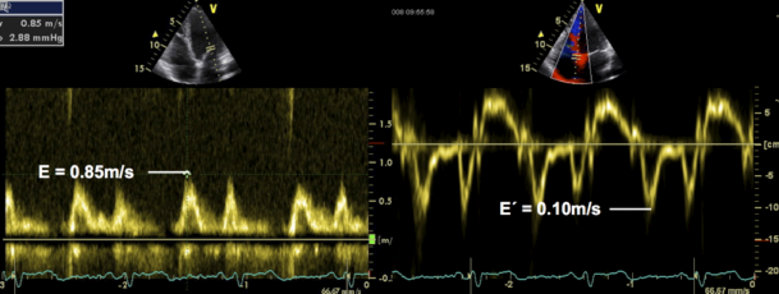
4.4 How to assess diastolic function | 123 Sonography

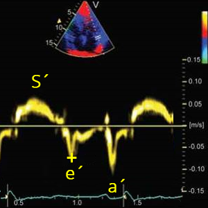
Tissue Doppler imaging (TDI) of the basal part of the right ventricle... |  Download Scientific Diagram
Tissue Doppler imaging (TDI) of the basal part of the right ventricle... | Download Scientific Diagram
4.4 How to assess diastolic function | 123 Sonography

Mitral annulus tissue Doppler imaging (TDI) | Download Scientific Diagram

Role of Tissue Doppler and Strain Echocardiography in Current Clinical  Practice | Circulation
Role of Tissue Doppler and Strain Echocardiography in Current Clinical Practice | Circulation

Tissue Doppler echocardiography - Wikipedia

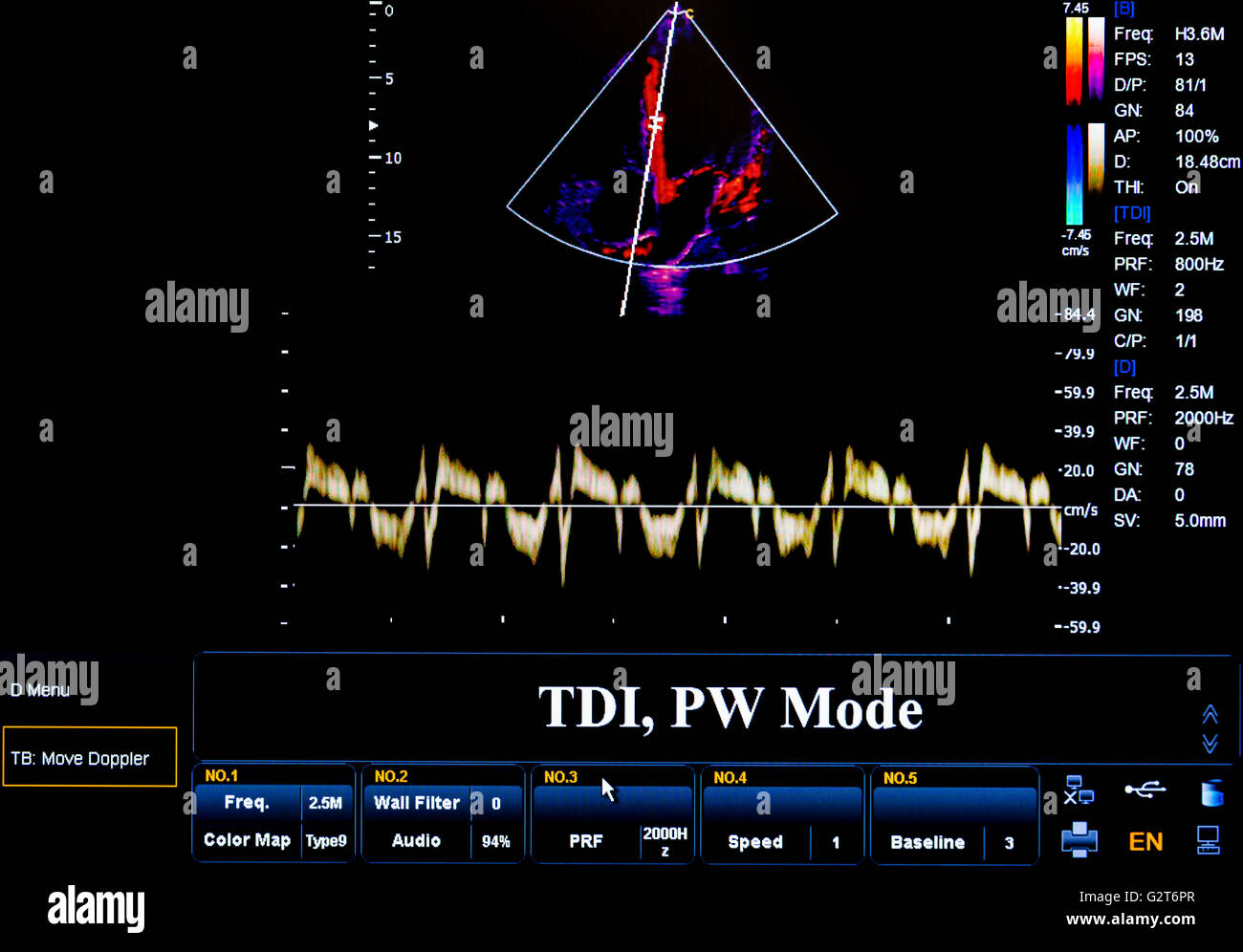
Modern echocardiography (ultrasound) machine monitor. Colour image. New  hospitl equipment. TDI, PW Mode Stock Photo - Alamy
Modern echocardiography (ultrasound) machine monitor. Colour image. New hospitl equipment. TDI, PW Mode Stock Photo - Alamy

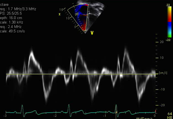
Tissue Doppler Imaging (TDI) velocities of tricuspid valve. After... |  Download Scientific Diagram
Tissue Doppler Imaging (TDI) velocities of tricuspid valve. After... | Download Scientific Diagram

Measurement of the atrial conduction time: PA-TDI is obtained using... |  Download Scientific Diagram
Measurement of the atrial conduction time: PA-TDI is obtained using... | Download Scientific Diagram
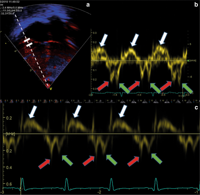
Tissue Doppler Imaging, Strain Imaging, and Dyssynchrony Assessment |  Thoracic Key
Tissue Doppler Imaging, Strain Imaging, and Dyssynchrony Assessment | Thoracic Key

Tissue Doppler velocity imaging and event timings in neonates: a guide to  image acquisition, measurement, interpretation, and reference values |  Pediatric Research
Tissue Doppler velocity imaging and event timings in neonates: a guide to image acquisition, measurement, interpretation, and reference values | Pediatric Research

Apical 4 chamber tissue Doppler | Pediatric Echocardiography

Tissue Doppler Imaging - an overview | ScienceDirect Topics

Tissue Doppler Echocardiography: Current Status and Applications - A  Practical Approach to Clinical Echocardiography 1st Edition
Tissue Doppler Echocardiography: Current Status and Applications - A Practical Approach to Clinical Echocardiography 1st Edition

Tissue Doppler echocardiography - Wikipedia

Tissue Doppler Myocardial Velocity Imaging in Infants and Children—a Window  into Developmental Changes of Myocardial Mechanics - Pauliks - 2013 -  Echocardiography - Wiley Online Library
Tissue Doppler Myocardial Velocity Imaging in Infants and Children—a Window into Developmental Changes of Myocardial Mechanics - Pauliks - 2013 - Echocardiography - Wiley Online Library
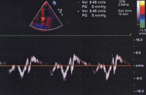
Example measurement of the PA-TDI interval. | Download Scientific Diagram

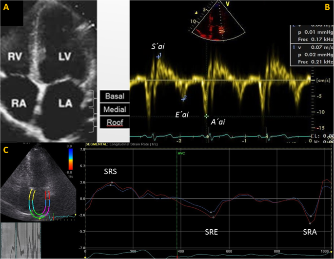
Tissue Doppler Imaging and strain rate of the left atrial lateral wall: age  related variations and comparison with parameters of diastolic function |  Cardiovascular Ultrasound | Full Text
Tissue Doppler Imaging and strain rate of the left atrial lateral wall: age related variations and comparison with parameters of diastolic function | Cardiovascular Ultrasound | Full Text